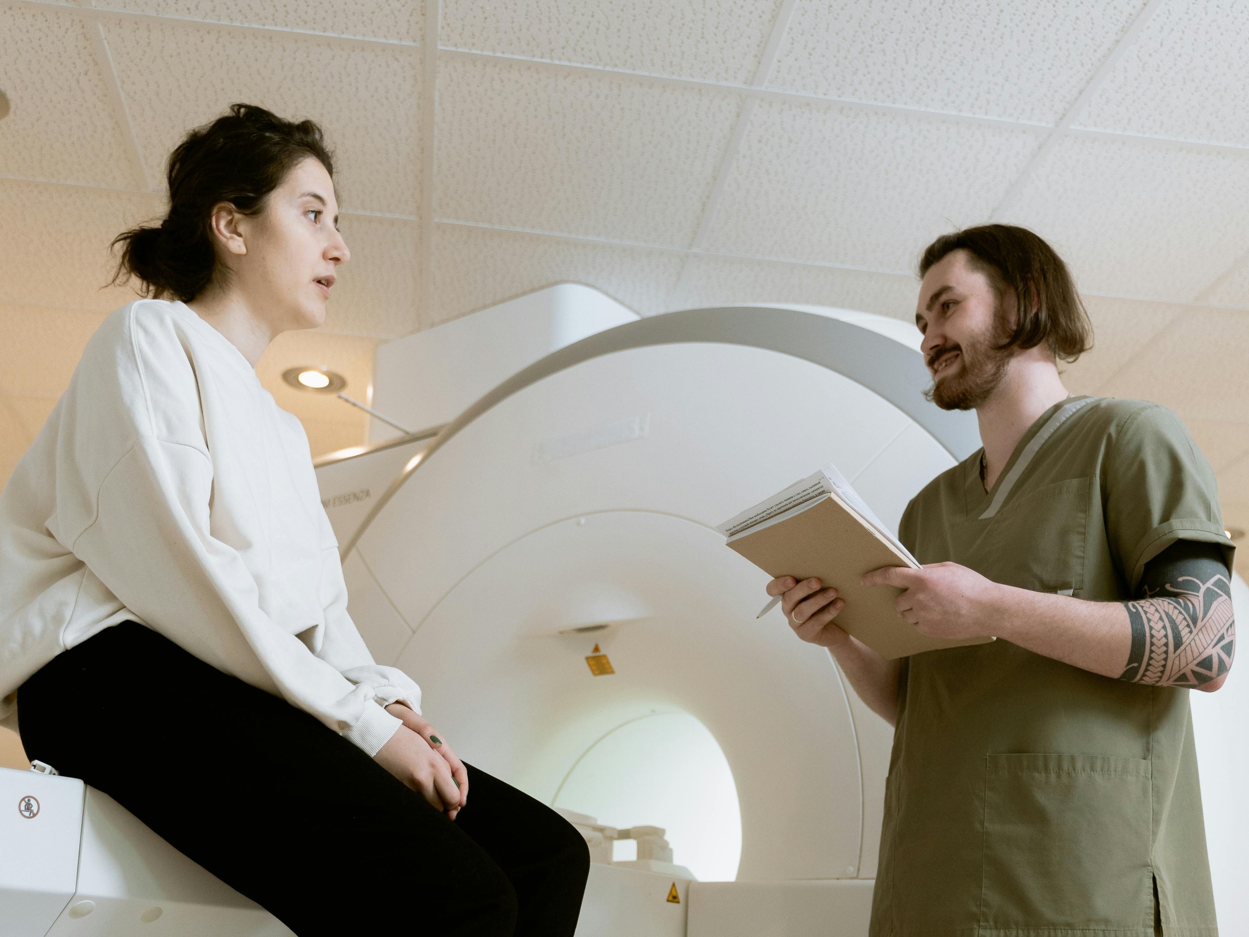
Video Blog: Cardiac MRIs

Empallo Heart-to-Heart
Welcome back to Empallo Heart-to-Heart! In this series, we explore common questions about heart health and cardiac care that don’t always get the time they deserve during a typical cardiology visit.
In today’s episode, Claire Beskin (CEO, Empallo, Inc.), Dr. Dana Weisshaar, MD (Medical Director, Empallo, Inc.), and Joe Lemay (CEO, Longevity Athlete) dive into a conversation about cardiac MRIs. We also spend some time revisiting cardiac CTs, building on what we covered in our previous episode.
We hope you find this helpful and engaging! Don’t forget, we’d love to hear your thoughts, so feel free to drop us feedback in the contact form on our website.
Check out the episode on Youtube. Enjoy!
____
[Transcript]
Claire Beskin: Hello and welcome to Empallo Heart-to-Heart. Today is April 1st, 2026 and we'll be talking about cardiac magnetic resonance imaging, more commonly called cardiac MRI. I'm Claire Beskin, founder and CEO of Empallo and I'm joined by Dr. Dr. Dana Weisar, medical director to Empallo, and Joe Lemay, Founder & CEO of Longevity Athlete. We started this series to discuss questions that frequently come up in cardiac care, but often don't get the time or space in a typical cardiology appointment to be fully explored. Our goal is to answer those questions that commonly come up and also spark new ideas that our listeners may not have thought about before. In a previous episode, we talked about cardiac CTS. Today we're focused on MRIs. Dr. Dana, could you give us a quick overview of MRIs, what they are, and when and why a cardiologist might order an MRI as opposed to a CT, for example?
Dana Weisshaar, MD: Sure. Thanks for asking. So, cardiac MRI. So, let's first start with MRI. So an MRI is a series of magnets that can take images and then process them into cross-sections or into images from different angles. So we can look at the heart in a way that is only possible because of the computing power that we have available. So, it takes these images of the heart that are quite detailed and allows the technician and the reader to manipulate the images to really get a three-dimensional look at all the cardiac structures. And of course, you can do an MRI of any part of the body, but today we're specifically talking about an MRI of the heart.
Claire Beskin: Along the same lines, what can a cardiac MRI show that an echo might miss?
Dana Weisshaar, MD: Well, an echocardiogram, which is also a two-dimensional image of the heart, is typically done what we call transthoracic, meaning we send sound waves through the body tissues from the skin and cross-sectioning in the heart. So, we can also get great images there, but we don't have as much fidelity when we get into the deeper tissues, the things that are further away from the chest wall. And so, the MRI can image finer details and deeper in because it's imaging the entire chest, not just the wedge that the ultrasound or the sound waves of an echo cardiogram can see.
Claire Beskin: Okay. I'm hearing that the MRI gives finer detail than the echo, right? Is there anything that the echo shows that the MRI would not?
Dana Weisshaar, MD: That's an interesting question. I think the answer is no. I think the MRI is going to see all of it. The reason we usually do echoes and not MRIs is because echos can be portable. They're much less expensive. They're much more available. So, ease, lower expense, and availability means we can have echocardiogram machines in every cardiology office, in every hospital, and everywhere. In fact, there are even mobile echo devices. So a company can come to your home for instance and do an echo.
Claire Beskin: Right. Okay makes sense. It sounds like cardiologists are putting together a lot of puzzle pieces between MRI, CT, echo. There's a lot of different imaging modalities and other diagnostics as well. And sometimes patients - and I - may have a hard time seeing how these pieces all fit together. So, it's helpful to see the bigger picture coming together.
Dana Weisshaar, MD: That's why we have all these different tools. They're tools for different things. It's as if I had a tool belt. And if all I have is a hammer, I'm going to hammer everything. But we have CT, echo, EKG, holters. But when we come to imaging, we can get imaging in a lot of different ways. So the cardiologist is going to choose whether it's a nuclear image or a CT image or an echo image or an MRI image based on the question being asked. So we want to put patients through the right tests to get the right answer. We don't want to put patients through a series of tests just because. And we don't want to put patients through more tests than they need to have done.
Claire Beskin: One of the first things patients usually ask is how to prepare for an MRI. For example, should they fast or avoid caffeine? Could you tell us a little bit about that?
Dana Weisshaar, MD: Sure. So, yes, you should avoid caffeine or smoking or really anything that is going to increase your heart rate right before the test because as we talked about with cardiac CT, we're talking about static imaging of a dynamic structure. So, the heart's always moving. In order to get good images, we have to gate the imaging in order to try to catch the heart kind of at the same spot at the same time. So, in general, a lower heart rate allows for better imaging because it's easier to gate it and capture it at that same spot. Whereas, if it's going faster, it's a little harder to gate. So avoid caffeine, avoid smoking, and generally you want to fast before this. Technically, if you are not getting contrast with your MRI, you could probably get away with having food beforehand, but it's generally a good idea to go in fasting in case something changes.
Claire Beskin: Right. So when you say in case something changes, does that mean that sometimes you get there and they determine that it would be better to use contrast dye even though that wasn't the original plan?
Dana Weisshaar, MD: Yes. So ideally the cardiologist when he or she is ordering the MRI will be specific about what they're looking for and then the technician and the performing cardiologist can adjust that to make sure they get the answer you need.
Claire Beskin: Right. So the question of whether to fast seems to come down to whether contrast dye is being used, but sometimes the patient may not know in advance with certainty whether contrast eye is going to be used. So as a best practice, if you can, try to fast, even if contrast dye wasn't specified in advance. Can you explain a little bit about when contrast is used, what it does, what patients should know about it, and how does this contrast dye differ from that used in other tests? For example, previously we talked about contrast dye in CT.
Dana Weisshaar, MD: Hey, that's a great question. So, it's a completely different contrast in MRI than in the CT. The CT contrast is an iodinated or has iodine in it. iodinated contrast. Whereas the MRI contrast is called gadolinium. They're both administered by IV, but they're not the same agent. In cardiac MRI, I would say most cardiac MRI does not need to have contrast. Contrast is used specifically to look for scarring, fibrosis generally in the heart muscle. And so what that looks like is the heart muscle looks black here. Let me show you an image. Heart muscle looks black and then the fibrosis will show up white. Can you see my screen?
Claire Beskin: Yes.
Dana Weisshaar, MD: All right. So here's an example. As I mentioned the muscle, so this is the left ventricle. So the muscle of the left ventricle is black. But here you can see: there's a white area. Or here's another example. The muscle is black and here's a stripe of white in the muscle. So that indicates fibrosis or scarring in the heart. As you can also see on this image, you can get quite a bit of detail about the size, the volume. You can calculate the volume based on the size and stacking of the images. And then you can see detail even much deeper into the chest like where the arteries - the pulmonary veins, actually - come into the left atrium. So this is detail that is too deep and too small for an echo to pick up but is easily seen on the cardiac MRI. I'll also point out over here, I mentioned that we can slice the images if you will in different ways through computer programming. And so here you're seeing the heart in what we call the long axis and then the computer can take cuts the other direction and give us a short axis. So we can see the heart from a variety of different angles to evaluate size and function.
Claire Beskin: You mentioned scarring in some of these images. How severe is this scarring?
Dana Weisshaar, MD: Okay, that's kind of a big question, so let me break it down. First is a disclaimer. I don't read cardiac MRIs for a living and so I can give basic information about scarring but I'm not going to be able to go into much subtlety about different types of scarring and how much gadolinium, late gadolinium enhancement, there is and things like that, which is a point that I should probably make clear - that while cardiologists today during their training are all exposed to cardiac MRI interpretation and ordering, not all cardiologists read these studies. They generally - because we don't do them that frequently and not every practice has access to a magnet - we generally, in a larger group of cardiologists, will have one or two cardiologists who specialize in cardiac MRI or who read all the MRI studies. They could be general cardiologists but they have a special interest in MRI. Similarly, in a large cardiology group, we would likely have one or two cardiologists who also read cardiac CT or we would have cardiologists who read nuclear medicine scans. Whereas any cardiologist can look at these images and understand them, but they're not necessarily experts in the interpretation of them. That said, here's what I can tell you. The amount of the myocardium that's involved is telling in terms of the extent of the fibrosis or inflammation. The location of the gadolinium enhancement is important. Is it a stripe in the middle of the myocardium, which is muscle, or is it a full thickness? This one's not even quite full thickness, but it's thicker. You can see it involves more of the muscle. And so the clinical correlation with that is if it's in a distribution that would suggest a prior heart attack or an MI, then we can say a patient has had a full thickness infarct or a partial thickness infarct. And that can speak to the viability of that part of the heart.
Dana Weisshaar, MD: Now, I'm getting kind of deep in the weeds, so let me back up a second and remind you that a heart attack is caused when a blood vessel supplying blood to the heart muscle closes, usually with a clot in it, and that chokes off the blood supply to the heart muscle. The heart muscle then becomes ischemic. Ischemia means not getting enough blood flow. And if it doesn't get blood flow long enough, the muscle can die. So it's as though someone's choking the heart muscle. They can choke you until you pass out. You're ischemic, but you're still viable, because if they stop choking you, you can recover. But if they choke you until you're dead, then you're not going to recover even if you stop choking. So the difference between viable and nonviable is that difference between is it ischemic or is it already dead - the cells. So the amount or the length of this stripe, the distribution of the stripe, speaks to which artery might be involved and how much of the heart was involved in a heart attack. And the thickness of the stripe might indicate that there's been some muscle cells that are dead, but not all of them in this area. So that's one of the things that the scarring - or what we call the LGE late gadolinium enhancement - tells us.
Claire Beskin: Coming back to a few of the questions I wrote down that our patients often ask. Another one that comes up is we talked about whether to avoid caffeine and avoid smoking before an MRI and the answer is generally yes. Avoid all those things. What about medications? Should people generally take everything as usual or are there some medications to avoid before an MRI?
Dana Weisshaar, MD: Generally, you should take all your medications as usual. If there's anything in particular that your cardiologist wants you to skip beforehand, they'll inform you, but there's no general rule that you have to.
Claire Beskin: All right, let's talk about wardrobe for a moment. My understanding is this is actually a pretty important question to address. So, Dr. Dana, what is the dress code for an MRI?
Dana Weisshaar, MD: So, the first and foremost thing to recognize is the M in MRI stands for magnet. And this is a big magnet. So, we definitely don't want any metals in the room because the magnet will attract them. So, you'll be leaving your watch, your keys, your jewelry all in a changing room, or you might even not want to wear any jewelry because you know you're going to have to take it off for the study.
Dana Weisshaar, MD: That actually brings up an interesting point too, which is there are some patients who can't safely have an MRI. Things like certain metallic foreign objects like shrapnel or welders if they get metal embedded may not safely have an MRI. So, it's not a test for everyone. Most surgical implants like hip replacements, valve replacements, those are fine for the MRI because they're non-magnetic metals. The thing to know since I mentioned valve replacements is that a valve replacement can cause shadowing in the MRI or the echo for that matter based on the properties of the valve replacement. But patients don't need to generally worry about that.
Dana Weisshaar, MD: The other question that oftentimes comes up is if I have a pacemaker, can I have an MRI? Because the answer used to be no. But device manufacturers are making what are considered MRI safe devices. They still require special care. Generally speaking, there will be a cardiologist at the MRI machine who will interrogate the device and may reprogram the device so that when you're having the MRI, your device is going to continue working without a hitch and then it gets interrogated after and maybe reprogrammed after. So, if you have an implantable device like a defibrillator or a pacemaker, certainly make sure that the technician knows about that and your cardiologist will usually have made special arrangements for managing that device before and after the MRI.
Dana Weisshaar, MD: But back to the question about clothing. So, you'll generally be asked to change into scrubs or a gown just to make sure you don't have a bra with an underwire, right? You just don't think about all the metal that's involved in your clothing. So, zippers, snaps, things like that are going to be no-no’s. So, if you're going to dress yourself for an MRI, wear something without any metal, zips, ties, underwires, things like that, but they'll generally have you change just to make sure.
Claire Beskin: One question that many people quietly wonder, but may not ask, is what does the MRI actually feel like? Is it uncomfortable or painful?
Dana Weisshaar, MD: That's an interesting question. It's not. I've had an MRI for other reasons, not a cardiac MRI, and I can honestly say it's not uncomfortable, except that it can be noisy. And it's a relatively small tube. It's a smaller tube than the CT scan tube. So, if you're sensitive to noises, then it might be somewhat uncomfortable. When I had my MRI, and most people when they have MRIs, the technician will give you earplugs because it's like a loud knocking and a lot of churning and chugging. It sounds - I can't tell you what it sounds like. It’s just a lot of knocking and churning sounds.
Dana Weisshaar, MD: If you're claustrophobic, it can be uncomfortable. So, if you are, then we'll talk to the technician about ways that it might be less claustrophobic. We're going to be imaging the heart. We're going to be imaging from kind of the upper part of the chest to the lower end of your rib cage. So, if it makes you claustrophobic to go in head first, maybe they can put you in feet first and up to here. Some people also who are super claustrophobic will get medications beforehand to kind of chill them out a little bit.
Claire Beskin: Okay. How long do people usually have to hang out in that tiny little tube listening to the MRI rock concert?
Dana Weisshaar, MD: So it depends on what studies or what your cardiologist is looking for. It can be 30 minutes or it can be 90 minutes. 90 minutes is a long time and that's generally when we're doing multiple different types of imaging, not just the straight non-contrast. So, if you're getting contrast, you'll generally get a non-contrast image and then get the contrast image after that.
Claire Beskin: 90 minutes. And I guess since there's a magnet, you probably can't bring a device in there. Can you bring a book or something - or what do you do in there for 90 minutes?
Dana Weisshaar, MD: Yeah, I don't know. I fell asleep during mine. So, but I can sleep anywhere, even with a big loud magnet banging around.
Joe Lemay: In mine, they had some entertainment options.
Dana Weisshaar, MD: Oh, that's good.
Claire Beskin: Tell us about your MRI experience, Joe.
Joe Lemay: A long time ago. It was for my foot and I've also had one for my shoulder. So yeah, it took more than a half hour sitting there and listening to the magnet do its thing.
Claire Beskin: What were the entertainment options?
Joe Lemay: They had a few radio stations I think available at the time. I kind of imagine now they might let you do a Bluetooth thing or something with your device, but who knows that there's probably different setups for different levels of fanciness and different facilities.
Dana Weisshaar, MD: Yeah, probably mostly just limited by the metal in any device.
Joe Lemay: Yeah, but they had speakers, so I imagine that now you could connect your phone to whatever speakers they have and that could work.
Dana Weisshaar, MD: But that's a great idea.
Joe Lemay: I don't really know.
Claire Beskin: This reminds me, I was recently catching up with an MIT computer science professor, Dr. Polina Golland who has been an adviser and mentor to us over the years and she was saying that in pediatric MRIs often infants and toddlers need to be put under general anesthesia to lie still enough throughout the MRI. And she runs a lab developing machine learning models focused on medical images. MRIs being one of the focus areas. And one thing that they've been developing is models that can sort of counteract motion. This may be a bad analogy, but it made me think about AirPods with noise cancelling technology, and they don't really eliminate the incoming sound, but they send out I think it might be called inverse sound waves or something like that that effectively neutralize the incoming sound. And so, I just thought about that conversation. I do wonder, Dr. Dana, when you see MRIs, to your knowledge, how often is motion an issue in MRIs?
Dana Weisshaar, MD: With the computer processing of the raw data, they can oftentimes clean up an image pretty well, but occasionally there'll be a disclaimer in the report that there's some motion artifact.
Claire Beskin: I went on Google and was searching for MRI reports because Dr. Dana, earlier you mentioned usually you would be looking at the report rather than the raw image. So I was on Google looking for an example MRI report and I ended up on Reddit, and I found an example report and I thought we might look at this together. So I'm going to share that in a moment.
Dana Weisshaar, MD: Let's take a look.
Claire Beskin: Okay. Can you guys see my screen?
Dana Weisshaar, MD: I can.
Claire Beskin: All right. We are in Reddit in Ask Cardiology. All right. And it says: cardiac MRI report. Is this normal? And there's some context down below:
“Hello, I'm a 25-year-old male. I recently did a cardiac MRI with contrast, without stress due to ongoing chest tightness and intermittent T-wave inversions on ECG. I've had multiple normal tests before (ECG echo, CT angiography, holter), but just wanted to rule out anything serious like myocarditis or cardiomyopathy. This is the first MRI I've done. And while the impression says no structural heart disease, I noticed that my myocardial mass index is slightly below the reference range. Just wondering, should I be concerned about the lower mass index? Does this suggest early cardiomyopathy or anything significant?”
Claire Beskin: Now, Dr. Dana, I know that this is not your patient and we're not treating this patient, but I did think this might spur some interesting conversation and also let our listeners and watchers see an example of what the report that comes to a cardiologist might look like. So, I wonder if you have any thoughts as you're looking at these questions posed by this Reddit user as well as this example MRI report.
Dana Weisshaar, MD: Sure. So the MRI report shows kind of what we talked about earlier that the MRI can be used to evaluate the volumes in the heart. Make sure the chambers are of normal size. Make sure that the wall thickness is normal and and then from that we can derive what the stroke volume is, which is how much blood is actually ejected with each beat of the heart. And we can calculate the ejection fraction which is, basically, no pump in the world is 100% efficient. So let's say you put 100 ml of blood in when it pumps you get 66.25 milliliters out with each contraction. So that's a normal ejection fraction. That's how we measure systolic function or the efficiency of the heart pump. And normal is 50% to 70% or 75%. So this person's quite normal. And you can see it looks very closely at the the heart structures, looks at the valves, and shows no myocardial edema that's what we talked about with the gadolinium enhancement and there's no scarring or fibrosis and so yes I agree this is a normal cardiac MRI.
Dana Weisshaar, MD: As to the question about should I worry about my low LV mass. As far as I'm aware, there's no correlation with cardiomyopathy or myocarditis related to low cardiac mass. It can be a sign of deconditioning. It can be a sign of malnutrition, but in the absence of those things, I wouldn't worry about that. So, I would say this is clearly a normal.
Dana Weisshaar, MD: What I will say is this patient's had a lot of different testing and it would be best for them to see a cardiologist. They probably already have. But to talk about microvascular dysfunction testing, which is a separate study in the cath lab.
Claire Beskin: Is that typically done after other tests are inconclusive?
Dana Weisshaar, MD: Yeah. So, the microvasculature, as it sounds, is small, right? So, so blood flows in, we talked earlier about how blood flows to the heart muscle. And so a CT angiography that this person mentions that he's had shows the larger blood vessels, but the smaller blood vessels can be dysfunctional and cause chest pain and EKG changes. And it's much harder to detect and much harder to confirm, which is why it likely needs to be done in a cath lab.
Joe Lemay: So I would ask a question.
Dana Weisshaar, MD: Sure.
Joe Lemay: Another one, I’m kind of curious. Do you think that the person who was concerned about Oh, did we even cover it here? I got it here. Okay, I will share.
Claire Beskin: All right. Joe is going to share his screen because there's another interesting Reddit question related to our previous episode about CTs.
Dana Weisshaar, MD: Ah
Claire Beskin: Specifically CT CACs. And so we're going to circle back to that topic. Joe, can you talk question a bit?
Joe Lemay: Sure. What I'm reading here is that this person got a calcium scan and kind of regrets getting the scan done in a way. There was some family history. But the calcium test came back with a 621, which terrified this person. And I guess in a nutshell, I'm wondering, do you think that it's good that this person got their CT calcium test, and do you think it's worth the stress, or do you think maybe I'll say the the last sentence that he or she asked is potentially, am I just overthinking this?
Dana Weisshaar, MD: Gotcha. Well, not having read the whole context, I'll say I think it's really good that this patient got the coronary calcium score because now they know that they have coronary artery disease. So it was there. Getting the test doesn't change whether or not the disease is present. What it changes is their awareness that, “Oh I have coronary disease.” Then the question is, “What am I going to do about it?”
Dana Weisshaar, MD: And so what they should do about it is take a statin first and foremost, not just to lower LDL cholesterol. Ideally, less than 100 is going to be our target if this patient's asymptomatic with their coronary disease, but it also stabilizes that plaque, that calcified plaque. The risk of a calcified plaque is that it can crack or break, and that's how a thrombus or a clot forms and causes a heart attack. So knowing that you have a high coronary calcium score means you need to take a statin. You need to modify any other risk factors for coronary disease. Don't smoke. Or stop smoking if you do. Know what your blood sugar or your A1C is and control your blood sugar. You can't change your family history. We talked about cholesterol.
Dana Weisshaar, MD: So the second part is that this person, if they have symptoms, may need a stress test to evaluate for ischemia or angina.
Dana Weisshaar, MD: And the third part is knowing that you have coronary disease, if heaven forbid you have sudden onset crushing subternal chest pain or some feeling of impending doom or symptoms, you're going to take it more seriously and go to the emergency room or call 911 because and you're going to say, "I have coronary artery disease and I have symptoms. I'm worried I'm having a heart attack." That is going to get that ball rolling to get you treated immediately and save the viability of the heart muscle.
Joe Lemay: That's good. I think I'll give more of the context here. I'll read it out:
“The calcium test came back with a terrifyingly high number, 621. Of course, I freaked out and spent the next few weeks thinking I was going to have a heart attack at any moment. I immediately shifted to a plant-based whole foods diet, cut out processed food, meat, and sugar completely. I was already in great shape as I've been playing soccer four to seven times a week in the last 10 years. I had no symptoms of any kind. By the way, this person is 52. Within a month of starting the plant-based whole food diet, a new blood test showed that I had reduced my LDL by a whopping 38%. Down to 105. Armed with these promising new numbers, I went to see the cardiologist who immediately did an EKG. Turned out to be totally normal. Prescribed Repatha twice monthly and baby aspirin daily and ordered a stress test. Took the stress test last week and I passed it with flying colors, getting to the end of level five. Even the guy who gave me the tests kept saying he was super impressed. So now I'm not sure what to make of anything. I feel like the calcium scores are potentially pointless given that a low score can produce a dangerous false sense of security. Plenty of people with low scores get heart attacks. And a high score can produce an equally dangerous, unnecessary high level of anxiety. Plenty of people with extremely high scores never have heart problems. Pre-calcium scan, I was super confident and not anxious about my heart health. At least until my mom's issues”
Joe Lemay: He found out his mom had some issues.
“Not paying the equivalent of an upscale gym membership fee every every month for Repatha and pretty much eating whatever I wanted. So potentially, am I overthinking this?”
Joe Lemay: So I guess my question to you is: do you think this person has improved their life or or chances of having a good life by having this information?
Dana Weisshaar, MD: Yeah. So um ignorance it might be bliss but it's not the best way to live a long healthy life. The fact that the cardiologist went directly to Repatha tells me that his starting LDL even though he did the right thing by going to a whole food plant-based diet and dropping it 38% that he still wasn't anywhere near the goal of less than 100 on the LDL. So probably has familial hyperlipidemia. So that said, knowing that his coronary calcium score is high is getting him to do the right things to improve his longevity. And it's not just longevity, it's living well into our later years. So having a heart attack may or may not kill you. If it doesn't, then it can leave you with heart failure or some disability later. So knowing that he has this, getting this treated now, while he's well, while he's able to play soccer, allows him to treat the problem before it gets worse and to stabilize that plaque and to reduce his risk of having an early heart attack. So living a natural life is fine. And living a natural life with super high cholesterol and/or coronary plaque ends in a heart attack or stroke at an early age. This way he's putting off that potential down the road.
Joe Lemay: And this CT score means he has heart disease?
Dana Weisshaar, MD: He has coronary artery disease. Yeah. Right. Given that he already has a coronary calcium score of 621, he has coronary artery disease.
Dana Weisshaar, MD: And so there are really three things I think that come from that. One is knowing you have coronary artery disease, get it treated, lower your LDL to less than a 100.
Dana Weisshaar, MD: And two, understand your other risks for coronary disease or or worsening coronary disease, controlling your sugars, don't smoke and and if you do smoke, stop smoking. You can't change your genetics. So managing the things you can do and then exercise, which is really important.
Dana Weisshaar, MD: And the third thing is now knowing that he has coronary artery disease. If he has sudden onset of crushing substernal chest pain or some other sense of impending doom or some feeling like that, he's going to know, “Oh, I might be having a heart attack.” Calling 911, heading to the emergency department immediately or within an hour of your chest pain or your discomfort and telling the ER doctor, “I have coronary artery disease and I have these symptoms” is going to trigger the activation of the process to look for and treat a heart attack in a more timely manner to save the viability of the heart muscle. Because having a heart attack and getting that artery open within 90 minutes saves muscle. So you have less of a scar from your heart attack than if you open the artery later, where the heart muscle's already died and now you've got a bigger scar.
Joe Lemay: I have a follow-up question. I heard is this true? I heard once, if you think you might be having a heart attack, one of the many things you should do is take an aspirin. Is that true?
Dana Weisshaar, MD:Yes. One of the things that we'll do in the emergency department is give you aspirin if you're on your way to the cath lab because the aspirin blocks thrombus formation which is clot formation. And the mechanism of the heart attack is that you rupture the hardened plaque - the calcium plaque - in your arteries and then that stimulates a clot formation within the artery and clogs the blood flow. So, the aspirin doesn't actually dissolve that clot. It just helps keep it from propagating or getting larger. Most of us don't have aspirin lying around the house. Don't stop at the store and get aspirin on your way to the ER. Just go to the ER. They'll give you aspirin there.
Joe Lemay: Does it matter, if you have it on your person? Is it good to take it? Like, does it matter if you take it a half hour earlier versus later or don't overthink it?
Dana Weisshaar, MD: Just don't overthink it. You can take it if you’ve got it on your person, but you know, don't let it get in your way of going to the emergency department.
Joe Lemay: That was great.
Dana Weisshaar, MD: Thanks. It sounds like we had enough for two blogs today.
Claire Beskin: All right, we've run over time. Today we covered MRI. We also sort of meandered back to CT, which we covered last time, but this was really interesting to bring in a real world example.
Claire Beskin: Thank you, Joe. Thank you, Dr. Dana. And looking forward to the next one.
Dana Weisshaar, MD: Great. Thanks for having me.
Joe Lemay: Great.
This content is for general informational purposes only and is not medical advice. Talk with your healthcare provider before making any decisions about your health.